
Case report
Ophthalmological manifestations of gyrate retinal atrophy. Case report
Manifestaciones oftalmológicas de la atrofia girata de retina. Informe de caso
Manifestações oftalmológicas da atrofia retiniana girata. Relato de caso
Carlos Alberto Pérez-Padilla1*  https://orcid.org/0000-0002-2873-9291
Zaihrys del Carmen Herrera-Lazo2 https://orcid.org/0000-0002-8015-0724
Christian Xavier Mata-Chango3 https://orcid.org/0009-0009-0212-6313
1Master's Degree in Comprehensive Child Care. First Degree Specialist in Ophthalmology. Subspecialist in Pediatrics Ophthalmology. Associate Professor. Universidad Regional Autónoma de Los Andes. Ambato General Teaching Hospital. Ambato, Tungurahua, Ecuador.
2Master's Degree in Comprehensive Child Care. First Degree Specialist in Pediatrics. Associate Professor. Universidad Regional Autónoma de Los Andes. Ambato General Teaching Hospital. Ambato, Tungurahua, Ecuador.
3Seventh-level medical student. Universidad Regional Autónoma de Los Andes. Ambato, Tungurahua, Ecuador.
*Corresponding author. Email:  makinario2013@gmail.com
ABSTRACT
Introduction: gyrate atrophy is a rare metabolic disorder of autosomal recessive inheritance. It is caused by a deficiency of the enzyme ornithine aminotransferase, which is inherited in an autosomal recessive pattern. Its chorioretinal findings are characteristic.
Objective: to present the case of a patient affected by gyrate atrophy, with emphasis on the diagnosis, the ophthalmological manifestations of the disease, and its treatment.
Case presentation: a 13-year-old female patient, of mixed race, residing in Ambato, Tungurahua province (Ecuador). She has a personal medical history of gyrate atrophy. She attended the consultation due to progressive decrease in visual acuity. On fundoscopic examination, hereditary choroidal atrophy, of a nonspecific nature was detected, extending from the posterior pole towards the periphery, in the form of circular rings (characteristic of the disease). She was prescribed a protein-restricted diet, with an emphasis on limiting arginine intake. The multidisciplinary follow-up protocol included quarterly monitoring of plasma amino acid levels to adjust dietary restrictions, as well as semiannual examinations for the early detection of secondary complications.
Conclusions: the diagnosis of gyrate retinal atrophy in this patient was based on the symptoms and the fundus examination. Genetic testing and the detection of elevated levels of systemic ornithine confirmed the diagnosis. Treatment based on reducing arginine intake lowers the risks of disease progression. Updated information on its diagnosis, ophthalmological manifestations, and treatment helps fill a gap in practical knowledge.
Keyworks: case reports; choroid; fundus oculi; gyrate atrophy; ornithine-oxo-acid transaminase; retina.
RESUMEN
Introducción: la atrofia girata es un trastorno metabólico raro, de origen hereditario autosómico recesivo. Se debe a la deficiencia de la enzima ornitina aminotransferasa, de herencia autosómica recesiva. Sus hallazgos coriorretinianos son característicos.
Objetivo: presentar el caso de una paciente afectada por atrofia girata, con énfasis en el diagnóstico, las manifestaciones oftalmológicas de la dolencia, y su tratamiento.
Presentación del caso: paciente femenina de 13 años, mestiza, residente en Ambato, provincia Tungurahua (Ecuador). Tiene antecedentes patológicos personales de atrofia girata. Acudió a la consulta por presentar disminución progresiva de la agudeza visual. En el examen de fondo de ojo se detectó atrofia hereditaria de coroides, inespecífica, desde el polo posterior hacia la periferia, en forma de anillos circulares (característicos de la enfermedad). Se le indicó un régimen dietético restrictivo en proteínas, con énfasis en la limitación de la ingesta de arginina. El protocolo de seguimiento multidisciplinario incluyó el monitoreo trimestral de los niveles de aminoácidos en plasma para ajustar las restricciones dietéticas, y la realización de exámenes semestrales para detectar precozmente complicaciones secundarias.
Conclusiones: el diagnóstico de atrofia girata de retina en esta paciente se basó en la sintomatología y el examen del fondo de ojo. El estudio genético y la detección de niveles elevados de ornitina sistémica posibilitaron confirmar el diagnóstico. El tratamiento basado en la reducción de la ingesta de arginina reduce los riesgos de agravamiento de la enfermedad. La información actualizada sobre su diagnóstico, manifestaciones oftalmológicas, y tratamiento, contribuye a llenar un vacío de conocimiento práctico.
Palabras clave: atrofia girata; coroides; fondo de ojo; retina; informes de casos; ornitina-oxo-ácido transaminasa.
RESUMO
Introdução: A atrofia girata é uma doença metabólica rara de herança autossômica recessiva. É causada pela deficiência da enzima ornitina aminotransferase, que é herdada em um padrão autossômico recessivo. Suas alterações coriorretinianas são características.
Objetivo: Apresentar o caso de uma paciente afetada por atrofia girata, com ênfase no diagnóstico, manifestações oftalmológicas da doença e seu tratamento.
Apresentação do caso: Uma paciente de 13 anos, mestiça, residente em Ambato, província de Tungurahua (Equador), apresentou histórico pessoal de atrofia girata. Ela procurou atendimento médico devido à diminuição progressiva da acuidade visual. O exame de fundo de olho revelou atrofia coroidal hereditária inespecífica, estendendo-se do polo posterior à periferia na forma de anéis circulares (característicos da doença). Foi prescrita uma dieta com restrição proteica, com ênfase na limitação da ingestão de arginina. O protocolo de acompanhamento multidisciplinar incluiu monitoramento trimestral dos níveis de aminoácidos plasmáticos para ajustar as restrições alimentares e exames semestrais para detectar precocemente complicações secundárias.
Conclusões: O diagnóstico de atrofia retiniana girata neste paciente foi baseado nos sintomas e no exame de fundo de olho. Os testes genéticos e a detecção de níveis elevados de ornitina sistêmica confirmaram o diagnóstico. O tratamento baseado na redução da ingestão de arginina diminui o risco de progressão da doença. Informações atualizadas sobre seu diagnóstico, manifestações oftalmológicas e tratamento ajudam a preencher uma lacuna no conhecimento prático.
Palavras-chave: atrofia girata; coroide; fundo de olho; retina; relatos de casos; ornitina oxoácido transaminase.
Received: 2025/12/16
Accepted: 2026/01/19
Published: 2026/02/05
It is important to have accurate diagnoses before scheduling refractive surgical interventions. To do this, the ophthalmologist must differentiate the diseases according to whether they are inflammatory, atopic, allergic, dystrophic or corneal degenerative in origin.(1) One must also take into account any alterations in the retina which may cause a sudden decrease in visual acuity from moderate to severe.(2)
Gyrate atrophy is a rare, autosomal recessive metabolic disorder of hereditary origin. It is due to the deficiency of the enzyme ornithine aminotransferase (OAT), encoded by the homonymous gene located at 10q26.13 with ten functional axons. This mitochondrial enzyme depends on pyridoxal phosphate (vitamin B6) and catalyzes the conversion of ornithine into glutamic acid and proline. Characteristic chorioretinal lesions are observed in patients.(3)
Enzyme deficiencies caused by mutations in the OAT gene cause high plasma concentrations of ornithine and progression of chorioretinal lesions characteristic of gyrata atrophy. Therefore, it is vital that patients assume an arginine-restricted diet to reduce plasma ornithine levels.(4)
It should be noted that chorioretinal atrophy is degenerative and progressive. Among the damages observable in the fundus examination, the chorioretinal circular atrophic areas are characteristic, clearly delimited and located mainly in the periphery of the retina. In elderly patients, the lesions are generally more numerous and larger, and affect the posterior pole of the retina.(4)
It is of the utmost importance that ophthalmology specialists are trained to diagnose and treat this rare disease, which causes serious complications during its evolution. Therefore, the objective of this article is to present the case of a patient affected by gyrata atrophy, with emphasis on the diagnosis, ophthalmological manifestations of the disease, and its treatment.
A 13-year-old female patient of mixed race who resides in Ambato, Tungurahua province (Ecuador). She has a known history of gyrate atrophy and he went to the ophthalmology outpatient clinic due to a progressive decrease in visual acuity.
History did not reveal any eye pain or other symptoms. Examination of the fundus (Fig. 1) revealed hereditary choroidal atrophy, nonspecific, from the posterior pole to the periphery, in the form of circular rings (characteristic of gyrate atrophy).
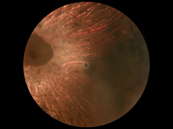
Fig. 1 - Color photograph of the fundus. Note the chorioretinal atrophic areas, clear in the periphery.
The patient's relatives expressed their consent to the publication of the case, by signing the informed consent document. The Ethics Committee of the hospital institution approved this scientific article.
The patient cooperated with specialists during the diagnostic procedures for her disease. Both she and her relatives were satisfied with the care received.
During physical examination of the patient, her mucous membranes were moist. In the examination of the respiratory system, normal breath sounds were heard. The respiratory rate was 20 breaths per minute. The heart sounds were rhythmic, of good tone and intensity; no murmurs were identified. The heart rate was 84 beats per minute, and the blood pressure was 118/79 mm Hg.
The upper and lower dental arches were complete, the tongue clean, the abdomen soft and depressible; no visceromegaly was detected. Normal bowel sounds were noted. The mental status exam showed that the patient was oriented in time and space; he did not present gait or language alterations, nor meningeal signs.
The patient received examination of the fundus and visual field perimetry. Peripheral degeneration of the retina, caused by gyrate atrophy, was verified and appeared progressive. The ophthalmological evaluation was comprehensive, to confirm the diagnosis and evaluate the progression of the disease.
In the fundus, multiple circular areas of chorioretinal atrophy were observed (Fig. 1), well defined, with confluent scalloped borders, located from the middle periphery to the posterior pole. The underlying choroidal vessels and relative papillary pallor were seen (Fig. 2).
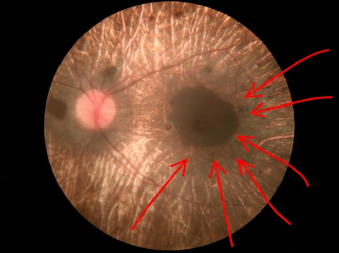
Fig. 1- Color photograph of the fundus. Note the atrophy of the retina, in the shape of a ring (indicated by the red arrows), characteristic of the disease.
The results of visual field and perimetry examinations revealed significant concentric narrowing of the visual field (tubular), with annular scotomas directly related to the areas of atrophy observed on fundoscopy. Peripheral vision was severely limited, and the patient retained only a small island of central vision.
Gyrate atrophy is caused by the functional deficiency of the OAT enzyme; therefore, the patient's treatment focused on reducing plasma ornithine levels to limit the progression of chorioretinal degeneration. A restrictive dietary regimen was indicated in proteins, with emphasis on limiting the intake of arginine (a direct precursor in the synthesis of ornithine), so as to avoid aggravation of ocular lesions and preserve residual vision.
As a complement, a therapeutic trial was initiated with pyridoxine hydrochloride at doses of 300 mg daily. The objective was to identify whether the patient was sensitive to vitamin B6, since this cofactor enhances the residual activity of the OAT enzyme, and makes it possible to significantly reduce hyperornithinemia.
At the same time, a multidisciplinary follow-up protocol was established for the patient, through quarterly monitoring of plasma amino acid levels to adjust dietary restrictions. From an ophthalmological perspective, every six months the patient will undergo computed perimetry and optical coherence tomography, to detect secondary complications such as cystic macular edema early, and to document the stability of the areas of atrophy observed in the diagnostic images.
Ornithine is deposited in various body tissues; the characteristic manifestations of this accumulation are myopia, cataracts, progressive chorioretinal atrophy, and macular changes. Hence, patients suffer from night blindness, reduced visual field, decreased central visual acuity, and blindness.(5) Dietary treatment restricted in arginine, or administration of vitamin B6 (precursor of the coenzyme pyridoxal phosphate), limit ornithine loading.(6)
The molecular mechanisms by which OAT deficiency causes retinal damage are not yet known.(6) However, it is known that due to the deficiency of this enzyme, plasma concentrations of the amino acid ornithine increase between 10 and 20 times compared to normal plasma levels; for which reason the ocular manifestations of the disease are attributed to it.(7)
The characteristic appearance of the fundus (choroideremia) is one of the lesions that can be seen early in patients. It consists of pigment concentrations in the retinal pigment epithelium, and peripapillary atrophy. Hence, in the advanced stage of the disease, the loss of the retinal pigment epithelium and choriocapillary originates confluent scalloped areas similar to those observed in later stages of gyrate atrophy.(8-10) These patients suffer from decreased visual acuity since childhood, associated with night blindness and poor peripheral vision.
Gyrate atrophy of the choroid and retina is secondary to OAT deficiency. This deficiency is not commonly diagnosed in childhood, so patients develop myopia and the characteristic retinal degeneration accompanied by hyperornithinemia. This is an indication that hyperammonemia, encephalopathy, and a biochemical profile characterized by low plasma ornithine, with citrulline and arginine, and increases in urinary excretion of homocitrulline and orotic acid (similar to a urea cycle disorder) appear in early childhood.(11)
Gyrate atrophy appears in the first decade of life, with a nearly equal proportion of males to females.(12) In most patients, the diagnosis is made or confirmed from the age of eight. The success of the treatment depends on its precocity, in a directly proportional ratio. The characteristic ophthalmological findings of atrophy appear from the age of six; approximately 30% of diagnosed patients develop macular edema, and 60% presenile cataracts.(13)
Variants of gyrate atrophy depend on the pathogenicity of the OAT gene in carrier patients. This means that homologous and harmful variants of OAT (c.G248A; p.S83) may appear, contributing to the progression of the disease.(14) If these varieties are repeated in each individual in a family, retinal imaging and metabolic analyses of pedigrees of consanguineous marriages can serve as guides for accurately diagnosing and treating the disease.
The most accurate diagnosis is based both on physical examination, particularly examination of the fundus (in which characteristic lesions due to plasma hyperornithinemia are observed), and on the detection of mutations in the OAT enzyme gene.(15,16) Early recognition and appropriate treatment of gyrate atrophy are essential to stop the sudden and sudden loss of visual acuity. Due to the frequent complications of this disease, peri- and postoperative adjuvant therapeutic modalities should be used, and the condition of patients should be monitored regularly.(17-19)
There is currently no effective gene therapy. The usual treatment consists mainly of restrictive dietary modifications of arginine (with pyridoxine, lysine, or creatine supplements). The aim is to reduce the intake of arginine as a precursor of ornithine.(20-22)
Preliminary ex vivo experiments have shown that red blood cells loaded with the OAT enzyme can metabolize extracellular ornithine at concentrations similar to those found in patients, both in plasma and in the blood supply. Therefore, it could be expected that a red blood cell-mediated treatment is feasible as a new therapeutic approach for this disease.(23)
This study was limited in terms of the lack of bibliography in Spanish and specific data on the incidence of gyrate atrophy, due to its rarity.
The diagnosis of gyrate atrophy in this patient was based on the symptoms and examination of the fundus. The genetic study and the detection of elevated levels of systemic ornithine made it possible to confirm the diagnosis. Treatment based on reducing arginine intake reduces the risks of worsening the disease. The research provided valuable scientific evidence on a rare condition, unknown in practice by many specialists. Up-to-date information on its diagnosis, ophthalmological manifestations, and treatment contributes to filling this gap in practical knowledge.
1. Albé E, Tassignon MJ. Eye diseases of importance in refractive surgery. Refractive Surgery. 2nd ed. Amsterdam: Elsevier; 2020. p. 132-145.
2. JF Salmon. Hereditary fundus dystrophies. In: Kanski: Clinical Ophthalmology. A systematic approach. 9th ed. Barcelona, Spain: Elsevier España, S.L.U.; 2021. p. 488-532.
3. Çavdarlı C, Şahlı E, Çavdarlı B, Numan M. "Regression of macular edema with topical brinzolamide and nepafenac alone and identification of a novel gyrate atrophy mutation." Arq Brasil Oftalmol [Internet]. 2020 [cited 2025 Oct 4];83(2):149-52. Available from: https://pmc.ncbi.nlm.nih.gov/articles/PMC11826744/pdf/abo-83-02-0149.pdf
4. Jalali H, Najafi M, Khoshaeen A, Mohammad-Reza M, Mahdavi M. "First report of c. 425-1G> A mutation in ornithine aminotransferase gene causing gyrate atrophy of the choroid and retina with hyperornithinemia." Eur J Ophthalmol [Internet]. 2021 [cited 2025 Oct 4]; 31(5):NP23-NP26. Available from: https://pubmed.ncbi.nlm.nih.gov/32418451/
5. Mejía-Alvarán M. and Ortiz-Zapata AI. Campimetric interpretation. In: Optic neuropathies from A to Z. Barcelona, Spain: Elsevier; 2024:121.
6. Montioli R, Bellezza I, Desbats MA, Borri-Voltattorni C, Salviati L, Cellini B. Deficit of human ornithine aminotransferase in gyrate atrophy: Molecular, cellular, and clinical aspects. Biochim Biophys Acta Proteins Proteom [Internet]. 2021 [cited 2025 Oct 4]; 1869(1):140555. Available from: https://www.sciencedirect.com/science/article/abs/pii/S1570963920302028
7. Elnahry AG, Tripathy K. Gyrate Atrophy of the Choroid and Retina. In: StatPearls [Internet]. Treasure Island (FL): StatPearls Publishing; 2024 Jan [cited 2025 Oct 4]. Available from: https://www.ncbi.nlm.nih.gov/books/NBK557759/
8. Balfoort BM, Pampalone G, Ruiter JPN, Denis SW, Brands MM. Extending diagnostic practices in gyrate atrophy: Enzymatic characterization and the development of an in vitro pyridoxine responsiveness assay. Mol Genet Metab [Internet]. 2024 Sep-Oct [cited 2025 Oct 4];143(1-2):108542. Available from: https://www.sciencedirect.com/science/article/pii/S1096719224004268
9. Elnahry AG, Elnahry GA. Rotated atrophy of the choroid and retina: a review. Eur J Ophthalmol [Internet]. 2022 [cited 2025 Oct 4];32(3):1314-1323. Available from: https://journals.sagepub.com/doi/10.1177/11206721211067333?url_ver=Z39.88-2003&rfr_id=ori:rid:crossref.org&rfr_dat=cr_pub%20%200pubmed
10. Labiano AT, Arroyo MH. Gyrate atrophy-like phenotype with normal plasma ornithine and low plasma taurine. GMS Ophthalmol Cases [Internet]. 2020 Feb 27 [cited 2025 Oct 4];10:1-5. Available from: https://pmc.ncbi.nlm.nih.gov/articles/PMC7113615/pdf/OC-10-04.pdf
11. Kaczmarczyk A, Baker M, Diddle J, Yuzyuk T, Valle D, Lindstrom K. A neonate with ornithine aminotransferase deficiency; insights on the hyperammonemia-associated biochemical phenotype of gyrate atrophy. Mol Genet Metab Rep [Internet]. 2022 Mar [cited 2025 Oct 4];16(31):100857. Available from: https://pmc.ncbi.nlm.nih.gov/articles/PMC9248225/pdf/main.pdf
12. Pauleikhoff L, Weisschuh N, Lentzsch A, Spital G, Krohne TU, Agostini H, et al. Clinical characteristics of gyrate atrophy compared with a gyrate atrophy-like retinal phenotype. Eur J Ophthalmol [Internet]. 2024 Jan [cited 2025 Oct 4];34(1):79-88. Available from: https://journals.sagepub.com/doi/10.1177/11206721231178147?url_ver=Z39.88-2003&rfr_id=ori:rid:crossref.org&rfr_dat=cr_pub%20%200pubmed
13. Palmer E, Stepien KM, Campbell C, Barton S, Iosifidis C, Ghosh A, et al. Clinical, biochemical and molecular analysis in a cohort of individuals with gyrate atrophy. Orphanet J Rare Dis [Internet]. 2023 Sep 4 [cited 2025 Oct 4];18(1):265. Available from: https://pmc.ncbi.nlm.nih.gov/articles/PMC10476330/pdf/13023_2023_Article_2840.pdf
14. Huang J, Fu J, Fu S, Yang L, Nie K, Duan C, et al. Diagnostic value of a combination of next-generation sequencing, chorioretinal imaging and metabolic analysis: lessons from a consanguineous Chinese family with gyrate atrophy of the choroid and retina stemming from a novel OAT variant. Br J Ophthalmol [Internet]. 2019 Mar [cited 2025 Oct 4];103(3):428-435. Available from: https://bjo.bmj.com/content/103/3/428.long
15. da Palma MM, Ku C, Igelman AD, Burr A, Shevchenko-Sutherland L. A possible ocular biomarker for response to hyperornithinemia in gyrate atrophy: the effect of pyridoxine, lysine, and arginine-restricted diet in a patient with advanced disease. Ophthalmic Genet [Internet]. 2023 [cited 2025 Oct 4];44(2):191-7. Available from: https://www.tandfonline.com/doi/10.1080/13816810.2022.2098986?url_ver=Z39.88-2003&rfr_id=ori:rid:crossref.org&rfr_dat=cr_pub%20%200pubmed
16. Merino-Diez MT, Soria-Prada C, Zamorano-Aleixandre M, Gonzalez-Lopez JJ. "Gyrate atrophy of the choroid and retina: Update on diagnosis and treatment. Arch Soc Esp Oftalmol (Engl Ed) [Internet]. 2024 [cited 2025 Oct 4];99(9):392-9. Available from: https://www.sciencedirect.com/science/article/abs/pii/S2173579424000719
17. Ozcaliskan S, Balci S, Artunay O. A Case of Foveoschisis Associated with Ornithine Aminotransferase Deficiency and Gyrate Atrophy. J Coll Physicians Surg Pak [Internet]. 2021 Nov [cited 2025 Oct 4];31(11):1354-6. Available from: https://www.jcpsp.pk/oas/mpdf/generate_pdf.php?string=dTJmR2pzTEpuSVQ2Tjh6T3N2cktzZz09
18. Berhuni M, Tıskaoğlu NS. Foveoschisis associated with gyrate atrophy in ornithine aminotransferase deficiency: A case report. Photodiagnosis Photodyn Ther [Internet]. 2023 Jun [cited 2025 Oct 4];42:103618. Available from: https://www.sciencedirect.com/science/article/abs/pii/S1572100023003459?via%3Dihub
19. Castaño-Silos JC, Sanchez-Guillen I, Almorín-Fernández-Vigo I, Jerez-Fidalgo M, Fernández-Vigo JI. Paravenous pigmented chorioretinal atrophy of unifocal and unilateral presentation. Arc Soc Esp Oftalmol [Internet]. 2023 Apr [cited 2025 Oct 4];98(4):233-7. Available from: https://www.sciencedirect.com/science/article/abs/pii/S0365669123000011
20. Ju Y, Zong Y, Li X, Gao F, Chang Q, Huang X. Mild Phenotypes of Gyrate Atrophy in a Heterozygous Carrier with One Variant Allele of OAT. Genes (Basel) [Internet]. 2024 Ago [cited 2025 Oct 4];15(8):1020. Available from: https://www.mdpi.com/2073-4425/15/8/1020
21. Pires-Franco de Oliveira BA, Vieira-Bussad M, Peyneau-Poncio L, Delpupo-Moro N. Choroidal gyrata and retina atrophy: update on diagnosis and treatment. Archives of Health, Curitiba [Internet]. 2025 [cited 2025 Oct 4];6(4):1-6. Available from: https://ojs.latinamericanpublicacoes.com.br/ojs/index.php/ah/article/view/3413/3117
22. Balfoort BM, Buijs MJN, Ten-Asbroek AL, Bergen AA, Boon CJ, Ferreira EA, et al. A review of treatment modalities in gyrate atrophy of the choroid and retina (GACR). Mol Genet Metab [Internet]. 2021 Sep-Oct [cited 2025 Oct 4];134(1-2):96-116. Available from: https://www.sciencedirect.com/science/article/pii/S1096719221007629?via%3Dihub
23. Pampalone G, Chiasserini D, Pierigè F, Camaioni E, Orvietani PL, Bregalda A, et al. Biochemical Studies on Human Ornithine Aminotransferase Support a Cell-Based Enzyme Replacement Therapy in the Gyrate Atrophy of the Choroid and Retina. Int J Mol Sci [Internet]. 2024 Jul [cited 2025 Oct 4];25:7931. Available from: https://pdfs.semanticscholar.org/d8cf/757a8c869b577e8e411f0e0ffea9a47f2822.pdf
Conflict of interest
The authors declare that there are no conflicts of interest.
Authors' contribution
Carlos Alberto Pérez-Padilla: conceptualization, research, methodology, project manager, resources, supervision, validation, visualization, writing of the original draft, writing, revision and editing.
Zaihrys del Carmen Herrera-Lazo: acquisition of funds, research, resources, supervision, visualization and writing, revision and editing.
Christian Xavier Mata-Chango: formal analysis, research, methodology, software, visualization and writing, proofreading and editing.
Funding
Ambato General Teaching Hospital. Ambato, Tungurahua, Ecuador.
 Esta obra está bajo una licencia internacional Creative Commons Attribution4.0/International/Deed.